
specialization
Fundus Imaging (Retina Scan)
Protect Your Vision with Advanced Fundus Imaging in Faridabad
- Home
- Fundus Imaging
Healthy vision depends on a healthy retina. Many serious eye diseases begin inside the retina without causing noticeable symptoms in the early stages. Fundus Imaging in Faridabad is one of the most advanced eye diagnostic tests used to examine the retina, optic nerve, and retinal blood vessels in detail. By capturing high-resolution images of the back of the eye, this examination helps ophthalmologists detect eye diseases early and monitor changes over time.
At Dr. Ruchi Eye Clinic, patients receive comprehensive retinal evaluations under the care of Dr. Ruchi Pahuja (MBBS, DOMS), who has more than 30 years of clinical experience in ophthalmology. Located in Sector 28, Rajendra Colony, Faridabad, the clinic uses advanced retinal imaging technology to support accurate diagnosis, personalized patient care, and an ethical treatment approach.
It is important to understand that Fundus Imaging in Faridabad is a diagnostic imaging test, not a treatment. It helps doctors detect, document, and monitor retinal and optic nerve conditions before significant vision loss occurs. Whether you need a Retina Scan in Faridabad, Digital Fundus Photography in Faridabad, or another Eye Diagnostic Test in Faridabad, early diagnosis plays an important role in protecting long-term vision.
What Is Fundus Imaging and Why Is It Important for Your Eye Health?
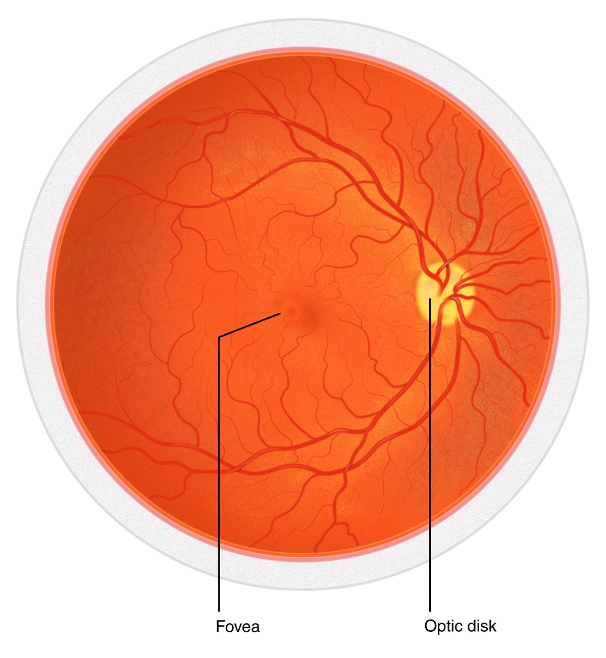
Fundus Imaging, also known as a retina scan or digital fundus photography, is an advanced eye imaging technique that captures detailed photographs of the retina, optic nerve, macula, and retinal blood vessels.
Unlike a routine eye examination, Fundus Imaging in Faridabad creates permanent digital images that allow your ophthalmologist to examine the inside of your eye in great detail. These images help detect very small retinal changes that may not be visible during a standard examination.
Fundus Imaging is commonly used to:
- Examine the retina in detail
- Evaluate the optic nerve
- Detect retinal diseases early
- Monitor diabetic eye disease
- Assess glaucoma-related optic nerve changes
- Document retinal abnormalities
- Support treatment planning
- Monitor long-term eye health
Many retinal diseases develop silently before affecting vision. Early imaging allows these conditions to be identified sooner, improving the chances of preserving healthy eyesight.
Patients requiring an Eye Diagnostic Test in Faridabad often benefit from fundus imaging because it provides accurate documentation of retinal health quickly and comfortably.
How Does Fundus Imaging (Retina Scan) Work?
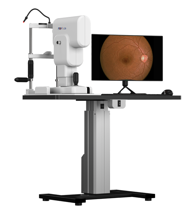
Fundus Imaging uses a specialized digital retinal camera to capture high-quality photographs of the back of the eye. The camera focuses through the pupil and records detailed images of important structures such as the retina, macula, optic nerve, and retinal blood vessels.
During the examination:
- High-resolution digital photographs are captured.
- The scan is completely non-invasive.
- No instrument touches the eye.
- Images are recorded within seconds.
- The photographs appear immediately for review.
- Your ophthalmologist carefully studies each image.
The examination is:
- Fast and comfortable
- Completely non-contact
- Highly accurate
- Suitable for routine monitoring
- Useful for long-term comparison
Because of its precision, Fundus Imaging in Faridabad has become one of the most valuable methods for evaluating retinal health and documenting changes over time. It is also widely used as Digital Fundus Photography in Faridabad for diagnosing and monitoring various retinal and optic nerve conditions.
Who Should Get Fundus Imaging in Faridabad?
Fundus imaging is recommended for people with eye symptoms as well as individuals who are at a higher risk of developing retinal disease.
Your ophthalmologist may advise Fundus Imaging in Faridabad if you:
- Have diabetes
- Have glaucoma
- Have high blood pressure
- Have Age-Related Macular Degeneration (AMD)
- Experience blurred vision
- Notice sudden vision changes
- Have a family history of retinal disease
- Need routine comprehensive eye examinations
- Require long-term retinal monitoring
Regular retinal imaging helps doctors identify changes before symptoms become severe. Early diagnosis often allows treatment to begin sooner and helps reduce the risk of permanent vision loss. At Dr. Ruchi Eye Clinic, every retinal image is carefully evaluated by Dr. Ruchi using advanced retinal imaging technology. Patients receive personalized recommendations based on their symptoms, medical history, and overall eye health. Whether you require a Retina Scan in Faridabad, Digital Fundus Photography in Faridabad, or another advanced Eye Diagnostic Test in Faridabad, early retinal evaluation supports accurate diagnosis and informed treatment decisions while helping protect your vision for the future.
Which Eye Diseases Can Fundus Imaging Detect?
The retina is one of the most important parts of the eye because it captures light and sends visual information to the brain. Even small changes in the retina or optic nerve can affect vision. Fundus Imaging in Faridabad helps ophthalmologists identify these changes early, often before symptoms become noticeable.
A detailed Retina Scan in Faridabad can help detect and monitor several eye conditions, including:
- Diabetic Retinopathy
- Glaucoma
- Age-Related Macular Degeneration (AMD)
- Retinal Vein Occlusion (RVO)
- Hypertensive Retinopathy
- Retinal Tears
- Retinal Hemorrhages
- Optic Nerve Disorders
- Macular abnormalities
- Changes in retinal blood vessels
Because many retinal diseases develop slowly, early diagnosis through Digital Fundus Photography in Faridabad allows timely monitoring and supports better treatment planning. It is important to remember that fundus imaging is a diagnostic imaging test, not a treatment. It provides valuable information that helps your ophthalmologist diagnose, document, and monitor retinal conditions accurately.
What Happens During a Fundus Imaging Test?
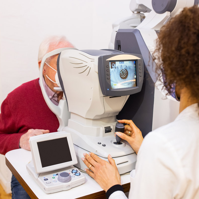
Most patients are surprised by how quick and comfortable the examination is. When you arrive for your appointment, the eye care team will first explain the procedure. In some cases, your ophthalmologist may recommend dilating your pupils with special eye drops to obtain a wider and clearer view of the retina.
During the examination:
- You sit comfortably in front of the retinal camera.
- Your chin and forehead rest on the support.
- You look at a small fixation target.
- The camera captures detailed retinal photographs.
- No instrument touches your eye.
- The images appear immediately for review.
The entire imaging process usually takes only a few minutes. If pupil-dilating drops are used, your vision may remain blurred for a short time afterward. Otherwise, most patients can return to their normal daily activities immediately. After the examination, Dr. Ruchi carefully reviews the retinal photographs and explains the findings in simple language.
Why Is Fundus Imaging Better Than a Routine Eye Examination?
A routine eye examination is important for checking vision and overall eye health. However, some retinal and optic nerve conditions require detailed photographic documentation that cannot always be achieved during a standard examination. Fundus imaging provides permanent digital records that allow doctors to compare retinal changes over time.
Compared with a routine eye examination, fundus imaging offers:
- High-resolution retinal photographs
- Early detection of retinal diseases
- Better documentation of retinal findings
- Monitoring of disease progression
- Accurate comparison during follow-up visits
- Improved treatment planning
- Detailed evaluation of the optic nerve
- Better long-term vision care
For example, early diabetic retinopathy, glaucoma-related optic nerve changes, or retinal bleeding may be detected and documented more effectively using Digital Fundus Photography in Faridabad.
At Dr. Ruchi Eye Clinic, advanced retinal imaging technology supports accurate diagnosis and comprehensive eye evaluations for patients of all ages. Located in Sector 28, Rajendra Colony, Faridabad, the clinic combines 30+ years of clinical experience, personalized patient care, and an ethical treatment approach to deliver reliable retinal assessments. Whether you need a Retina Scan in Faridabad, Digital Fundus Photography in Faridabad, or another Eye Diagnostic Test in Faridabad, detailed retinal imaging helps your ophthalmologist make informed decisions that support better long-term eye health.
Why Is Fundus Imaging Important for People with Diabetes and High Blood Pressure?
Diabetes and high blood pressure can damage the small blood vessels inside the retina long before vision changes become noticeable. Regular Fundus Imaging in Faridabad helps detect these changes early and allows treatment to begin before serious complications develop.
Retinal imaging is especially important because it can identify:
- Diabetic retinopathy
- Hypertensive retinopathy
- Retinal bleeding
- Swollen retinal blood vessels
- Optic nerve changes
- Macular abnormalities
Since these diseases often progress silently, routine retinal screening plays an essential role in preventing avoidable vision loss. Patients with diabetes or hypertension should never wait for symptoms before scheduling retinal examinations.
Frequently Asked Questions
Fundus Imaging is an advanced retinal imaging test that captures detailed photographs of the retina, optic nerve, and retinal blood vessels. Fundus Imaging in Faridabad helps diagnose, document, and monitor retinal diseases while supporting accurate treatment planning and long-term eye care.
Yes. Fundus Imaging is commonly referred to as a Retina Scan in Faridabad because it captures detailed digital photographs of the retina. The test allows ophthalmologists to evaluate retinal health, identify abnormalities, and compare changes during future visits.
No. Fundus Imaging is a completely non-contact and painless procedure. No surgery, injections, or radiation are involved. As an advanced Eye Diagnostic Test in Faridabad, it is comfortable, safe, and suitable for routine monitoring of retinal health.
People with diabetes, glaucoma, high blood pressure, AMD, blurred vision, or a family history of retinal disease may benefit from regular retinal imaging. Your ophthalmologist may also recommend Digital Fundus Photography in Faridabad as part of a comprehensive eye examination.
Yes. Fundus Imaging in Faridabad helps identify diabetic retinopathy, glaucoma-related optic nerve changes, retinal hemorrhages, hypertensive retinopathy, and several other retinal conditions. It provides valuable photographic documentation that supports early diagnosis and long-term monitoring.